
The five fingers on our hand are divided into three bones called distal, middle and proximal phalanx. Which are likely to break following an injury.
As a diagnostic radiographer I see so many fractured fingers during x-ray procedures and usually my patients ask me questions about the recovery timeline.
For this article I rely on my knowledge as a radiographer and scientific studies on the subject matter to answer your questions..
The aim is to reassure you about the progression of this fracture.
Happy reading!😀
All references at the end of this article, let’s meet in the comments section🙏
Last updated: Feb 2025. Written by Juliet Semakula, diagnostic radiographer.
Disclaimer: Amazon affiliate links: complete disclosure in the legal notices
▶️Basic Anatomy of fingers:
Our fingers are made of small bones called ‘phalanx’ named as proximal, middle and distal phalanx as demonstrated in the image below.
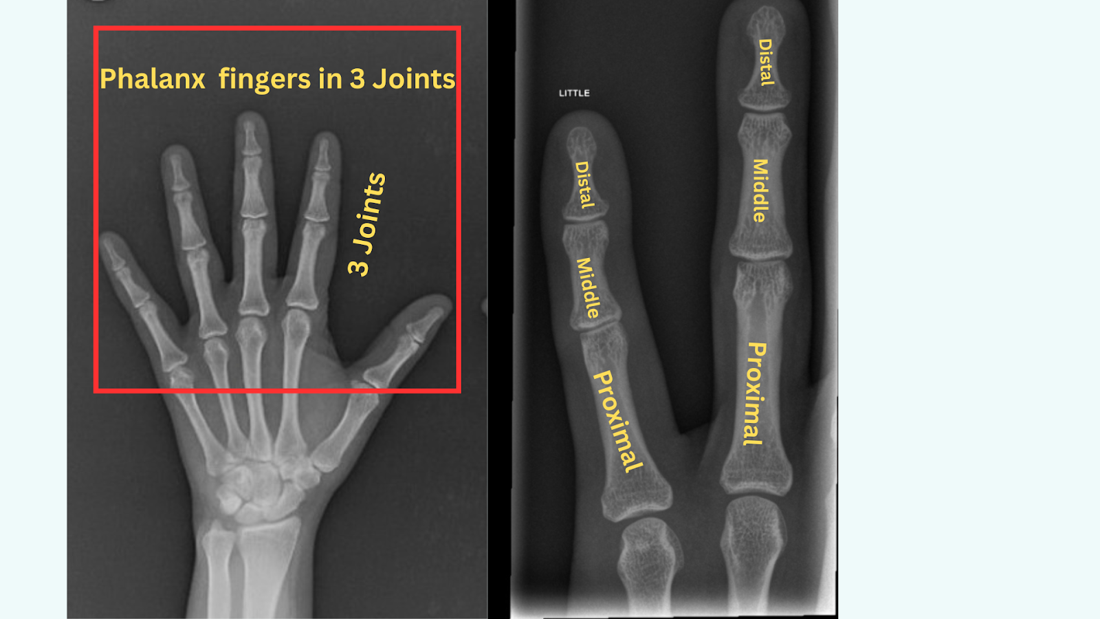
X-ray image showing hand distal, middle and proximal phalanx fingers
▶️What are the most common spots and names of phalanx fracture to the finger
There are different kinds of phalanx finger fractures. And treatment can be different for each type.
It is generally an orthopaedic surgeon who determines which treatments to pursue based on the type of fracture.
Here are some of the names of phalanx finger fracture injuries: transverse, oblique, bicondylar, volar, dorsal base, vertical, radial base and tuft.
The image below clears shows the different locations of phalanx finger fracture.
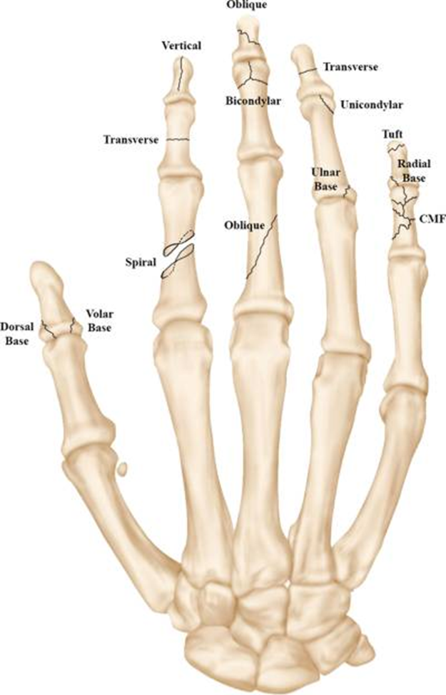
Image from: Moura et al 2022
X-ray images showing examples of distal, middle and proximal phalanx finger fractures

X-ray images showing the oblique middle phalanx fracture ,distal tuft phalanx fracture and oblique proximal fracture of the finger
▶️How do you treat a phalanx fracture?
Whichever treatment method your doctor decides to use to treat your phalanx injury.
Usually, the overriding goal is to restore anatomy and impart enough stability to allow for early motion of your finger.
There two types of treatment options normally used will depend on the type of fracture you have got.
1️⃣Conservative management
🟠 Buddy-taping or finger splint
Usually the injured finger is buddy-taped to the adjacent digit finger to control rotation during the healing process or a splint is used to align the fracture. Different types are available on Amazon or at any pharmacies
This allows early motion and decreases joint stiffness
🟠A thermoplastic splint may be used for some people to protect the joint and promote healing.
During this process a few close monitoring is done until clinical healing is noted, usually the splint is removed at 3 weeks
2️⃣Surgical option:
If your fracture is unstable, involving a joint, tendon or ligament it may need surgical management to re-align the fracture.
I have seen two techniques used in theatre while providing x-ray for surgeons during the operation.
🟠Kirschner wire (K-wire) fixation techniques
Metal wires are used to align the bone using x-ray to guide the surgeon. Fixation under the extensor tendon is avoided to minimise friction
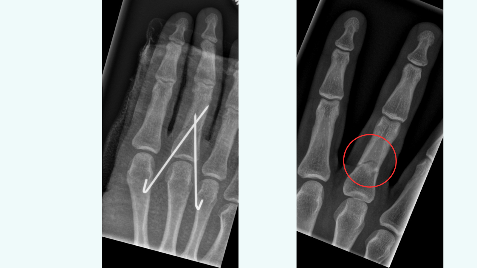
Image on the left shows Kirschner wire (K-wire) fixation methods for proximal phalanx fracture. And on the right x-ray images showing k-wire removed.
This is a trans-articular technique where the K-wire crosses the metacarpophalangeal joint prior to crossing the fracture site.
🟠And there is a periarticular technique where the pins start at the radial and ulnar from the base of the proximal phalanx and cross the fracture site.
▶️What is the outcome or result from the surgical option
Studies on how effective the 2 parallel trans-articular K-wires inserted radial and ulnar to the extrinsic extensor tendon showed
🟢Closed pinning minimises additional soft tissue injury and allow for early motion
🟢According to some studies, a good overall range of proximal interphalangeal joint motion is noted at 96% after treatment (Lögters,2017)
🟢Most phalangeal fractures treated by plate and/or screw fixation achieved a good range of motion more than or equal to 210 degrees (Kozin,2000)
The K-wires are normally removed at 3 to 4 weeks after the operation.
Screw fixation
Sometimes when your fracture is oblique and spiral screw fixation will be used
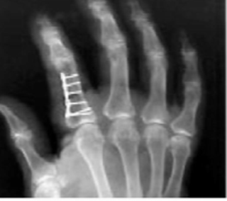
Postoperative PA film obtained after open reduction and internal fixation with a mini-condylar blade plate secured on the lateral border Image from: Kozin, 2000
If screws and plates are used to fix the fracture, they normally remain in place
🔴Sometimes they cause finger stiffness related to adhesions between the hardware and the extensor mechanism
According one comparison study between trans-articular and periarticular pinning methods showed neither fixation method to be superior (Safi,2012)
▶️What are the possible complications of a finger phalanx fracture after treatment?
🔴Finger stiffness.
🔴Infection is always a concern after fracture fixation
🔴Non-union or malunion this when there is a poor healing of the bone leading to finger deformity, stiffness and pain.
🔴Plate prominence and loosening
🔴Tendon rupture can occur in 57% of these fractures (kozin, 2000) rare thou.
🔴Scarring of the soft tissues can occur in the skin, around tendons, or in joint capsules.
🔴Complex regional pain syndrome (CRPS)
▶️Is a phalanx finger fracture serious?
Over the years of my radiographer career, I have heard these questions among patients I see in x-ray.
They usually wonder if it was a waste of time for them to seek medical advice for a fractured finger.
The truth is, you will only know whether you have fractured your finger bone or not by seeking medical treatment.
An injury like a broken finger or thumb might seem small, but that doesn’t mean it’s unimportant
Your finger may look normal to you because you can still have a normal range of movement which is possible even with a broken finger.
Stop being embarrassed or not sure if you should seek medical treatment, get your injury checked by a professional.
For one, your finger will be examined and an x-ray requested to determine if you have fractured it or not.
However small fingers can fracture and not show, I do x-ray so many fractured fingers every day at work.
Reasons why it is important:
▶️What happens if you leave a fractured finger untreated?
⚫Pain around the injured finger will not go away by itself.
⚫Your finger may end up deformed if not attended to in time.
⚫Stiffness and limited range of movement.
⚫Swelling around the finger
⚫Bruising and purple colour when blood is not circulating well to the hand.
⚫Tenderness to touch and redness around the affected area.
⚫Numbness and limited movement require immediate attention to avoid damaging the nerves tendons and ligaments.
Remember the chances of successful treatment increase the sooner your treatment starts. It is advisable to always see a doctor within a week of the injury for quick recovery.
▶️How long does it take for a fractured phalanx of a finger to heal?
Summary: it usually takes 6 to 12 weeks for the distal, middle and proximal phalanx fingers to heal whether conservative or surgical.
Whether you have fractured a distal, middle or proximal finger, the consolidation normally takes 6 weeks. In some cases, it may take longer to heal.
You will have a follow up and a post check x-ray to see how your fracture is healing.
Most fractures I have seen heal irrespective of the method of fixation.
But I have also seen a small percentage of delayed consolidation, poor consolidation and non-union in post check x-rays.
Your physical therapist or your doctor or surgeon will be able to tell you what is appropriate or not in your case in case of delayed healing.
However, most patients I have seen regain their functionality and are able to return to their normal activities.
Here are some proposed timeline of finger healing I have observed in patients.
| Weeks since injury | Stages of healing |
| Less pain | Few weeks |
| Finger Swelling and bending | 6 weeks to a Couple of months |
| Strapping or wearing a splint | 0 to 3 weeks |
| Strapping or splint removal | 3-6 weeks or more |
| Complete Healing | 6 -12 weeks, some could take a year of discomfort. |
| Return to normal activities | 6 to 12 weeks |
| 12 weeks | If you still have pain & swelling, always consult the fracture care team for advice. |
Proposed healing time of phalanx fingers ,it may be more less in children
▶️Will I need Physiotherapy for my fractured finger?
The aim of a physiotherapy session is to help you gradually resume the range of movement in your fingers and be able to go back to your previous activities.
To achieve this a physiotherapy will help you with:
⚫Therapy for scar mobilisation.
⚫Tendon gliding
⚫Joint mobilisation
Your physio will guide you with finger exercises to help you gain joint release to restore motion.
Some people decide to exercise at home following the physio instructions, it will depend on what you want.
We have come to the end of this article, any questions and experience let us meet in the comments section.
▶️Can you speed up healing and consolidation of a phalanx finger?
There are no drugs, devices or physiotherapy techniques that can speed up bone consolidation. This is a natural process that can take several weeks.
You can only help the process by:
⚫Following your doctors’ instructions of resting and taking it easy with your finger.
⚫Reduce or stop smoking because it is known to interfere with the healing process.
⚫Consider taking vitamin D calcium supplement or consuming more nutritious foods if you are deficient.
⚫Be patient you finger will function normally like before.
We have come to the end of this article below are some of the references I have used.
Wishing you a quick recovery!🙋
Other articles that might interest you.
⚫ Living with non-union fracture.
⚫Why fractured bones hurt more at night.
⚫ Dislocated finger, treatment and recovery
⚫ A broken thumb: recovery and rehabilitation
📚Source:
Moura SP, Meulendijks MZ, Veeramani A, Szapary H, Gomez-Eslava B, Hoftiezer YAJ, Chen NC, Eberlin KR. Epidemiology and Fracture Patterns of Traumatic Phalangeal Fractures.PlastReconstrSurgGlobOpen.2022Aug 4;10(8):e4455. doi: 10.1097/GOX.0000000000004455. PMID: 35936823; PMCID: PMC9351885.
Lögters TT, Lee HH, Gehrmann S, Windolf J, Kaufmann RA. Proximal Phalanx Fracture Management. Hand (N Y). 2018 Jul;13(4):376-383. doi: 10.1177/1558944717735947. Epub 2017 Oct 27. PMID: 29078727; PMCID: PMC6081790.
Faruqui S, Stern PJ, Kiefhaber TR. Percutaneous pinning of fractures in the proximal third of the proximal phalanx: complications and outcomes.JHandSurgAm.2012Jul;37(7):1342-8.doi: 10.1016/j.jhsa.2012.04.019. PMID: 22721457.
Kozin SH, Thoder JJ, Lieberman G. Operative treatment of metacarpal and phalangeal shaft fractures. J Am Acad Orthop Surg.2000Mar-Apr;8(2):111-21.doi:10.5435/00124635-200003000-00005. PMID: 10799096.
Oxley PJ, Fin Hodge W. Functional Hand-Based Splint in the Treatment of Metacarpal Fractures. Plast Surg (Oakv). 2023 Nov;31(4):350-357. doi:10.1177/22925503211042867. Epub 2021 Dec 23. PMID: 37915347; PMCID: PMC10617463.