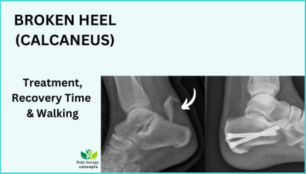
Have you been diagnosed with a fracture of the calcaneus (heel bone), or are you looking for information about treatment and recovery tips. You are in the right place.
In this article, I address the most common questions asked from patients I see in x-ray with heel injuries.
To provide answers, I draw upon my experience as a diagnostic radiographer as well as extensive research in medical scientific publications. All sources cited are at the end of this article.
Happy reading!😀
Have any questions, remarks, or experience to share? Feel free to use the comments section at the end of the article!🙏
Last updated: June 2024. Written by Juliet Semakula, diagnostic radiographer.
Disclaimer: no affiliate links.
Basic anatomy
The calcaneus or heel bone is a complex shaped bone located below your ankle and extending to the muscle to your feet.
The heel bone is a very important bone that supports you as you walk, and it also connects your calf muscles to your foot that allows you to push off as you take a step forward.
Anatomy of a calcaneus
▶️What does a calcaneus fracture look like with or without displacement?
Heel fractures are rare injuries, during my years of practice I have seen a few heal injuries, but when they do happen, they can be potentially unbearable injuries.
Overview: Different types of calcaneus fractures.
1️⃣Intra-articular fractures. These are considered the most serious type of heel fractures because they involve damage to the cartilage between the joints
2️⃣Avulsion fractures. This where a fragment of bone is avulsed away from the calcaneus from the pulling from the Achilles tendon or another ligament.
3️⃣Multiple fracture fragments. This is also known as a crushed heel injury and is more common with higher impact injuries such as a fall from height or automotive accident.
4️⃣Stress fractures. While most calcaneal fractures are caused by trauma, a calcaneal stress fracture can result from overuse or repetitive trauma like high volume of running or jumping.
According to medical studies extra articular fractures account for 25% of calcaneal fractures. These typically are avulsion injuries of the calcaneal tuberosity from the Achilles tendon.
Calcaneal or calcaneus fractures account for 50-60% of all fractured tarsal bones.
Less than 10% present as open fractures where the bone has pierced the skin.
Sources: Statpearls 2024
Treatment will depend on the type of fracture and the same guidelines are followed whether displaced or not.
▶️What causes calcaneus fractures.
Calcaneal fractures most commonly occur during high energy impact such as
🟢Fall or jumping into a hard surface from height and any injury to the foot and ankle.
🟢Penetrating trauma and twisting/shearing events may also cause injury to the heel.
🟢Stress fractures to the calcaneus may occur with overuse or repetitive use, such as running.
🟢Diseases such as diabetes and osteoporosis may increase the risk of heel fractures.
Most patients with calcaneus fractures are young male, between the age of 20-39 due to the nature of the injury.
▶️How serious is a calcaneal fracture?
Calcaneus fractures can be significantly serious injuries and you should seek medical treatment if you think you have a fracture.
Most of these injuries cause the bone to flatten, widen, and shorten causing you the following symptoms:
🟢You will feel diffuse pain around the affected site.
🟢Swelling or edema which may last for months depending on the damage of the bone and cartilage.
🟢You will not be able to bear weight on the heel.
🟢Bruising and tenderness.
🟢There may be associated injuries of the Achilles tendon which usually is an indication of a calcaneus injury.
▶️Diagnosis of a calcaneus fracture
Your doctor will send you to x-ray or CT for imaging and your medical report will likely describe the type of injury you have got.
This will depend on the location and type of injury to the bone, it could be minimally with a few fracture lines and bone fragments, displaced with a slight gap or intra or extra articular fracture.
X-rays and CT images show clearly the outline of the fracture which helps with treatment
▶️What are the treatment options of calcaneus fractures?
The treatment you get will be determined by the type of calcaneus fracture you have and your general health. It could be through a conservative or surgical method which all leads to healing.
1️⃣Conservative or orthopaedic treatment: Splint, cast.
To treat a minor calcaneus stress fracture, your doctor will recommend the following methods:
You limit or avoid putting weight on your foot for four to 8 weeks or more.
You will also be prescribing a boot, cast or splint for 8 to 10 weeks to keep your foot in one position while the fracture heals.
Heel fracture treated conservatively with a boot or cast: Image showing a foot wear which can help to protect the heel from touching the feet
In some cases, there may be no specific immobilisation, but you are advised to be cautious with ankle movements.
Weight-bearing is often delayed 8-12 weeks or more depending on the degree of comminution and progression of healing on routine post x-rays.
▶️Can a calcaneus fracture heal without surgery?
Most calcaneus fractures I have seen heal without surgery, using the conservative methods we have discussed above.
However, some patients do complain of pain even after treatment, questioning if the fracture is healing well.
When you think your fracture is not healing as it should always consult your doctor.
It could be an infection, or it might be healing in a position that causes discomfort or trouble with walking or wearing shoes.
Some medical studies do not recommend surgical methods for closed, displaced or intra-articular fractures of the calcaneus due to the higher complication rate of infection and discomfort. It has been spotted that sometimes it causes removal of hardware metal in 11% of postoperative patients (Griffin 2014)
While most calcaneus fractures can heal without surgery, sometimes healing can take longer than anticipated and it can come with lifelong complications such as
♦️Arthritis.
️ ♦️Stiffness and pain in the joint frequently develop.
♦️Sometimes the fractured bone fails to heal in the proper position.
♦️Decreased ankle motion and walking with a limp due to collapse of the heel bone and loss of length in the leg.
♦️Patients often require additional surgery or long-term or permanent use of a brace or an arch support to help manage these complications.
2️⃣Calcaneus Surgical and placement of plates or screws
The decision to move forward with surgery will be based on fully informed consent with you due to the risks and expected benefits of surgery.
Before surgery soft tissue swelling should be resolved completely which normally takes 10-14 days to avoid further complications. Buckley 2004
Open surgery: Your calcaneal will be reconstructed to something close to its original form a plate or screws will be used to stabilise the fracture site.
Percutaneous minimally invasive technique will be used where a small insertion will be made then a plate will be used to slide under the skin and fixed to the bone with a few screws.
▶️After surgery complications are common and may include:
♦️Compartment syndrome.
♦️Osteomyelitis.
♦️Infection can occur in mild cases which can be treated with antibiotics given to you by your doctor.
♦️️ Wound healing problems caused by weak circulation to the heel’s soft tissues, the surgical site may not heal properly and may require additional wound care or skin grafting.
♦️️Non-union here the bone simply doesn’t heal. If this is the case, the use of a bone growth stimulator (non-invasive) may be beneficial. In more severe cases, this may require additional surgery to help the bone resume normal healing.
♦️Subtalar arthritis in the joints which could cause you chronic pain. If this occurs, at some point you may need to undergo an additional procedure to fuse the affected joints.
Clare 2016.
In summary, complications surrounding the treatment of calcaneus fractures remain a challenge for orthopaedic surgeons.
However, you should understand that the goal of operative management is to restore natural anatomy to maximise function and lifespan of the joint.
▶️Physical therapy rehabilitation after calcaneus fracture will be divided into two.
🅰️ Post operation rehabilitation once the healing process is underway. Here the physio will guide you on how you can gradually start to allow your leg and heel to bear weight and some motion to avoid stiffness.
🅱️Rehabilitation care during the healing process will aim at reducing any complications and reduce pain.
After your operation your foot will be placed into an extremely well-padded posterior splint to protect your feet.
You will be given personalised advice. Here in the health sector (NHS), the PRICER rules is followed which is abbreviated in full as
P-Protect:
After your operation your foot will be placed into an extremely well-padded posterior splint to protect it.
R-rest:
Try to rest your feet, no weight-bearing after surgery for a few weeks.
I-ice:
Apply ice to help reduce pain and swelling.
C-compression:
wearing a compression stocking to reduce swelling is sometimes advisable.
E-elevation:
After your operation you will be advised to elevate your leg for the first few weeks to help reduce swelling and prevent the risk of wound complications.
R-rehabilitation:
You will be referred to a physical therapy to help you with motion exercises to the subtalar joint and ankle to avoid stiffness as soon as possible.
This brings me to answering this common question.
▶️Can you walk on a fractured calcaneus and is it normal to have a lot of pain?
A calcaneus fracture is very painful. You can’t walk or put weight on your foot. You’ll also have a lot of swelling, bruising and tenderness.
Which is normal, the pain tends to decrease over the course of days, usually 21 days or weeks.
You need to give yourself time to heal before you think of putting pressure on your heel. When you start weight bearing you may need to adopt your movement with the help of walking aid.
You should try walking with reduced weight-bearing on the heel using appropriate splint or footwear and you may use crutches. A leg footwear support without putting body weight on the heel.
Very often in the United Kingdom and United States you will be advised to be immobilised and non-weight bearing for at least 6 weeks or more depending on your injury.
However, for certain patients If your injury is minor, such as a crack in the bone with little muscle damage, you may be able to resume normal activities from 3 to 4 months after surgery.
If your fracture is severe, however, it may take from 1 to 2 years before recovery is complete.
▶️How long does it take for a broken heel bone to heal?
When our bones break, an automatic repair usually occurs straight away where new bone tissue will be created without the need for any specific intervention.
Your job is to help assist this process by following your doctors’ instructions and changing your lifestyle if you are a smoker, stop during the healing process and have good nutrition to support the healing process.
| Weeks since injury | Rehabilitation plan |
| 0-6 weeks | · Wear the boot all the time when walking, you can take it off in bed.· Use the crutches to take some of the weight off your foot. |
| 6-8 weeks | · Gradually discontinue using the boot and elbow crutches.· Try walking around the house without them first.· Wear the boot when walking longer distances outdoors. |
| 6-12 weeks | · Disappearance of foot swelling. |
| 8-12 weeks | · Fracture should be largely united (healed).· Gradually resume normal activities as pain allows.· Heavier or more strenuous tasks, including long walks, may still be difficult and cause discomfort and swelling at this stage. |
| 12+ weeks | · Symptoms will continue to improve over the next few months.· If you are still experiencing significant pain or stiffness, please contact your doctor for further advice. |
| 4 weeks to 3 months | · Returning to work |
| 6 months to 2 years | · Full functional and muscular recovery. |
Healing time after fracturing your heel, whether you have undergone surgery or not.
▶️How long should you wait to drive after a calcaneus fracture?
In general, it is advisable to wait until you can bear weight on the leg, and you feel fully recovered.
What does the United Kingdom driving regulation policy say about this?
You can only return to driving when:
♦️You are no longer using your boot.
♦️You can walk comfortably.
♦️You can perform an emergency stop pain free.
And you must tell the driving agency (DVLA) if you think you will be unable to drive for more than 3 months because of a broken limb if you fail to do so you can be fined up to 1000 pounds.
What does the United States and Canadian regulation driving policy say?
In the United States and Canada, there are no specific nationwide regulations regarding driving after a calcaneus fracture. Each state may have its own guidelines or recommendations.
Generally, individuals are advised to wait until they have recovered enough mobility, strength, and confidence to safely operate a vehicle.
We have come to the end of this article. I hope I have answered some of your commonly asked questions.
I wish you a very quick recovery!🙋
📚Sources:
Park ES, Choi Y, Lee J, Park SH, Lee HS. Calcaneal fracture: results of earlier rehabilitation after open reduction and internal fixation. Arch Orthop Trauma Surg. 2021 Jun;141(6):929-936. doi: 10.1007/s00402-020-03575-4. Epub 2020 Aug 11. PMID: 32780200.
Rammelt S, Swords MP. Calcaneal Fractures-Which Approach for Which Fracture? Orthop Clin North Am. 2021 Oct;52(4):433-450. doi: 10.1016/j.ocl.2021.05.012. Epub 2021 Jul 29. PMID: 34538353.
Smitaman EE, Davis M. Hindfoot Fractures: Injury Patterns and Relevant Imaging Findings. Radiographics. 2022 May-Jun;42(3):661-682. doi: 10.1148/rg.210167. Epub 2022 Mar 11. PMID: 35275783.
Clare MP, Crawford WS. Managing Complications of Calcaneus Fractures. Foot Ankle Clin. 2017 Mar;22(1):105-116. doi: 10.1016/j.fcl.2016.09.007. Epub 2016 Dec 20. PMID: 28167056.
Griffin D, Parsons N, Shaw E, Kulikov Y, Hutchinson C, Thorogood M, Lamb SE., UK Heel Fracture Trial Investigators. Operative versus non-operative treatment for closed, displaced, intra-articular fractures of the calcaneus: randomised controlled trial. BMJ. 2014 Jul 24;349:g4483. [PMC free article] [PubMed] [Reference list]
Treasure Island (FL): StatPearls Publishing; 2024 Jan
Buckley RE, Tough S. Displaced intra-articular calcaneal fractures. J Am Acad Orthop Surg. 2004 May-Jun;12(3):172-8. [PubMed] [Reference list]
Image: https://orthoinfo.aaos.org/en/diseases–conditions/calcaneus-heel-bone-fractures/
